

Cone Beam Imaging in Delray Beach, FL
Cone beam imaging in Delray Beach, Florida, provides a precise, three-dimensional view of teeth, jaws, and facial structures to support accurate diagnosis and treatment planning. At Sepe Dental, this technology helps map details that standard two-dimensional X-rays can miss, improving clarity for implants, root canals, orthodontics, and complex extractions. The scan is quick, comfortable, and designed to deliver detailed information while keeping radiation exposure as low as reasonably achievable.
Cone Beam Imaging Explained
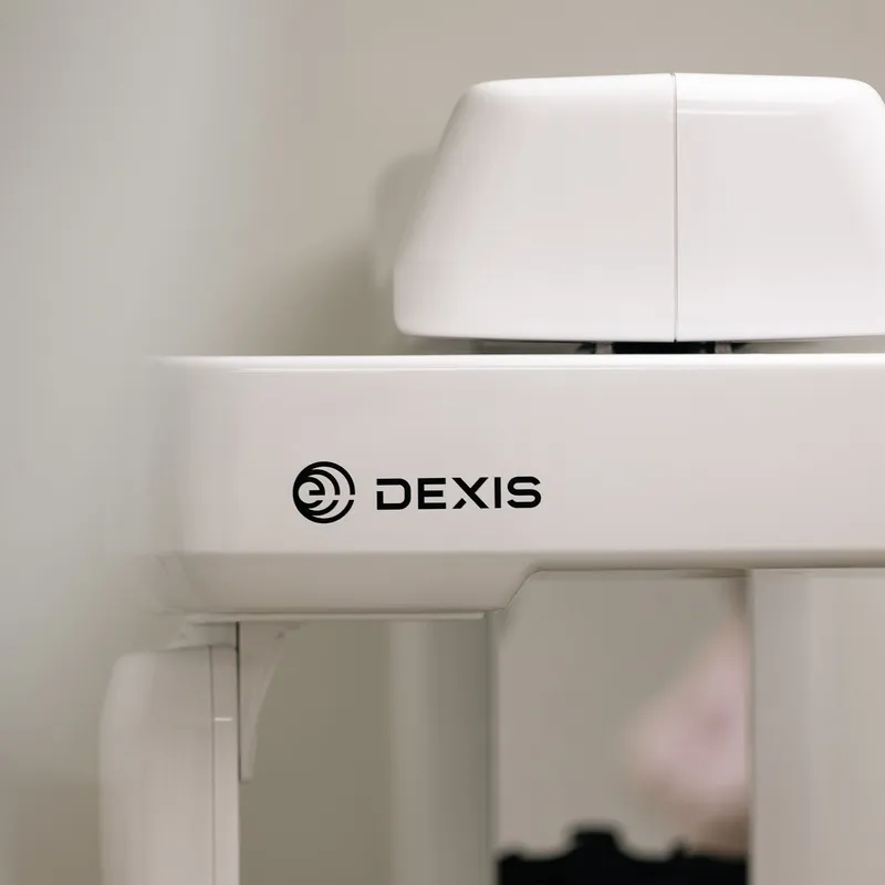
CBCT uses a cone-shaped X-ray beam and a rotating scanner to capture hundreds of images in seconds. Specialized software reconstructs those images into a 3D model of your teeth, bone, nerves, and sinuses.
With this view, our dentist can evaluate bone quality for dental implants, pinpoint canal anatomy for root canal therapy, and locate impacted teeth with precision. Compared with medical CT, dental cone beam imaging typically has a lower radiation dose tailored to the area of interest.
Benefits of Cone Beam Imaging
- ✓Comprehensive 3D detail for accurate diagnosis and planning.
- ✓Targeted views that focus on the region being treated.
- ✓Improved implant planning with assessment of bone height and width.
- ✓Clear visualization of nerves and sinuses to reduce surgical risks.
- ✓Enhanced evaluation of root canals, fractures, and hidden infections.
- ✓Efficient scans that are quick, comfortable, and easy to complete.
Need Advanced Imaging?
Schedule your cone beam scan for precise diagnosis and treatment planning.
How Cone Beam Imaging Works
The process is straightforward:
Assessment and Need
Our dentist determines if a 3D scan will change or improve your care.
Positioning
You stand or sit while your head is gently stabilized to limit movement.
Scanning
The machine rotates around your head for 10–20 seconds to capture images.
Reconstruction
Software builds a 3D model, often available within minutes.
Review
Our dentist studies the views to guide diagnosis and treatment steps.
Your Cone Beam Imaging Experience
Most scans take less than a minute from start to finish, and the exposure itself lasts only seconds. No special preparation is required, though you may be asked to remove earrings, glasses, or removable dental appliances. The scan is painless and noninvasive, and there is no downtime afterward.
Radiation dose varies by machine and field of view but is generally lower than a medical CT of the same region. Pregnant patients should inform the dental team so timing and protection can be considered. Images may reveal findings beyond teeth, such as sinus issues or bone variations, which can be helpful for comprehensive planning.
We Can't Wait to See You!

Frequently Asked Questions
Cone beam CT creates a 3D model of your teeth, jaws, and surrounding structures. It is used when a three-dimensional view will improve diagnosis or guide treatment, such as for implants or complex root canals.
Regular X-rays provide flat, two-dimensional views. CBCT produces a 3D image, revealing depth and orientation. This added detail can improve accuracy for implant placement and surgical planning.
CBCT uses ionizing radiation, but the dose is typically lower than a medical CT of the same area. The scan is prescribed only when the expected diagnostic benefit outweighs the small risk.
Yes. CBCT evaluates bone height, width, and density, and maps nearby structures like nerves and sinuses. This supports safer placement and helps select the right implant size and position.
No special preparation is needed. Removing metal items near the face helps reduce image artifacts. The scan is quick, comfortable, and you can return to normal activities immediately.
Our dentist reviews the 3D images, often the same day. The detailed views inform diagnosis and allow a clearer explanation of your treatment choices and next steps.
Related Dental Care and Patient Resources
Explore related treatment options, meet your dentist, review payment information, or plan your visit to Sepe Dental.